research
The Heart's Smallest Network Kept It Beating
Mouse experiments separated two tiny cardiac-neuron populations: one sustaining baseline rhythm, the other coordinating the response to stress.
Summary
Mouse experiments separated two tiny cardiac-neuron populations: one sustaining baseline rhythm, the other coordinating the response to stress.
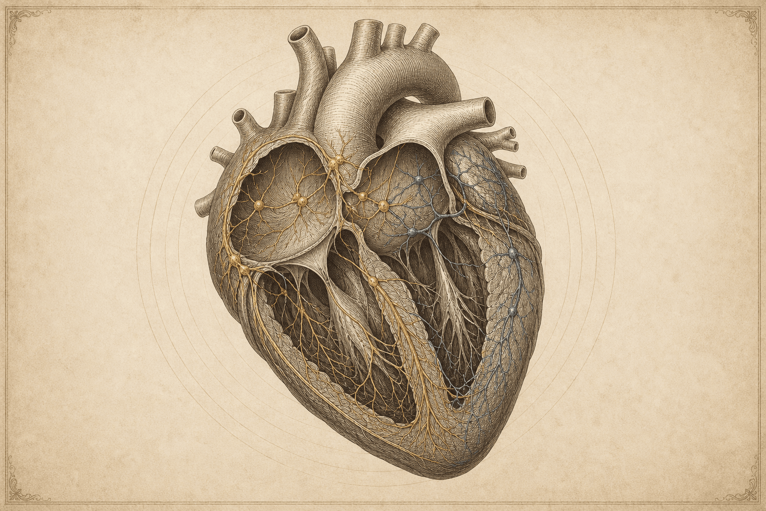
Yale researchers identified two major populations in the heart's intrinsic neural network. Removing Npy-positive neurons caused mouse hearts to stop, while Ddah1-positive neurons proved important when the animals faced physiological stress. Together the cells make up less than 0.01% of heart tissue. The Cell study establishes distinct functions in mice; it does not yet show that manipulating these neurons is a safe treatment in people.
Why it matters
Mouse experiments separated two tiny cardiac-neuron populations: one sustaining baseline rhythm, the other coordinating the response to stress.
Limits and context
- The Cell study establishes distinct functions in mice; it does not yet show that manipulating these neurons is a safe treatment in people.
Key claims
Mouse experiments separated two tiny cardiac-neuron populations: one sustaining baseline rhythm, the other coordinating the response to stress.
Qualification: The Cell study establishes distinct functions in mice; it does not yet show that manipulating these neurons is a safe treatment in people.
Evidence: source-2026-07-31-002
Sources
- Yale School of Medicine via Newswise: Cardiac neurons are essential for heart functionYale School of Medicine · secondary reporting
Corrections
No corrections have been recorded for this story.